All published articles of this journal are available on ScienceDirect.

Environmental Transport, Bioaccumulation, and Health Impacts of Micro- and Nanoplastics: An Integrated Review
 Mrityunjay Kumar2
Charu Arora3
Mahavir Parshad4
Durgesh Dixena5
Indrajit Mahto6
Ashu Goshisht7
Authors Info & Affiliations
Abstract
Introduction
Micro- and Nanoplastics (MNPs) are pervasive environmental contaminants with increasing concern for human health and ecosystems. Their small size, persistence, and physicochemical diversity permit bioaccumulation, long-range transport, and trophic transfer covering aquatic, terrestrial, and atmospheric systems.
Methods
This review comprehensively integrates old to recent literature on the sources (both primary and secondary), physicochemical properties (surface chemistry, shape, size, charge, and chemical composition), transport (through soil, water, air, food webs, etc.), exposure routes (dermal, ingestion, and inhalation), detection and characterization techniques, toxicity (in vertebrates, invertebrates, aquatic organisms, etc., and plants), challenges, and mitigation of toxicity of MNPs.
Results
Worldwide, evidence-based studies have reported the presence of MNPs in soil, water, food, air, and within organisms and plants. Physicochemical properties of MNPs strongly influence their distribution, exposure, and interaction with organisms. For example, most studies reported higher toxicity of nanoplastics than microplastics. Nanoplastics trigger oxidative stress, inflammation, genotoxicity, and autophagy, and cross various biological barriers, such as the lymphatic system and the gut lining. Microscopy, spectroscopy, thermal, and hybrid techniques play a crucial role in the detection and characterization of MNPs. All studies emphasized controlling MNPs' exposure, distribution, and toxicity by adopting effective, diverse strategies and raising awareness worldwide.
Discussion
Plastic pollution (especially micro- and nanoplastics) is one of the major challenges today. Moreover, micro-and nanoplastics are ubiquitous. They are heterogeneous, adsorb various other pollutants and microbes, and transport them in different ecosystems. Their physicochemical properties allow them to enter biological systems and reach the liver, kidneys, heart, and the lymphatic and gut systems. They cause oxidative stress, inflammation, DNA damage, mitochondrial damage, autophagy, necrosis, and apoptosis. Moreover, nanoplastics cause higher toxicity than microplastics. Hence, there is an urgent need for advanced and standardized detection and characterization techniques.
Conclusion
MNPs pose a significant threat to the health of vertebrates, invertebrates, aquatic organisms, and plants. They cross multiple biological barriers, disrupt numerous organs/systems, and trigger apoptosis. There is an urgent need for advanced detection techniques to overcome MNPs. Integrated mitigation approaches are essential for effective regulatory action.
1. INTRODUCTION
Plastics, especially Micro- and Nanoplastics (MNPs), have recently emerged as a major concern for terrestrial and aquatic environmental health and safety owing to their ubiquitous presence across ecosystems (Thompson et al., 2004). The constantly increasing production of plastics, their widespread use in everyday life, and inadequate, half-hearted waste management practices have led to their extensive environmental circulation, even into the Arctic regions (Andrady, 2011; Gigault et al., 2018). While the collection and recycling of bulk plastic waste may appear feasible, environmental weathering processes fragment plastics into MNPs, which are far more difficult to detect, monitor, and remove. There are two sources of MNPs: primary sources (intentionally produced powders, paints, inks, microbeads, toothpastes, abrasive blasting, biomedical devices, etc.) and secondary sources (existing from the breakdown of macroplastics like bottles, bags, packaging films, tyres, road wear, synthetic fibers, etc.) (Cózar et al., 2014). Notably, MNPs have also been reported in human waste and indoor air. Indoor air has been reported to contain substantial levels of airborne MNPs (Nizzetto et al., 2016; Bergmann et al., 2019). Alarmingly, MNPs have also been detected in the feces and blood clots of newborns, as well as in maternal breast milk (Liu et al., 2023; Wehrli et al., 2025). In marine ecosystems, MNPs have been reported in fish, turtles, crustaceans, and other higher organisms. Lower trophic levels, such as plankton, shellfish, and suspended algae, were also reported to internalize the MNPs. The diversity of morphologies, polymer types, sizes, colors, and shapes further complicates their environmental assessment.
Exposure of organisms to MNPs through inhalation, dermal contact, and ingestion elicits immune responses. However, nanoplastics, being smaller in size (≤ 1 μm) than microplastics (≤ 5 μm), possess greater efficiency in penetrating cells and interacting with various biological macromolecules, including proteins, DNA, mitochondria, and lysosomes (Trevisan et al. 2022; Lehner et al., 2019; Feng et al., 2023). Then, they trigger cellular damage, oxidative stress, genetic mutations, inflammation, and other health risks (Teuten et al., 2009). In addition, the biodistribution, biocompatibility, and toxicity of surface-altered NPs nanoplastics may differ appreciably from those of pristine NPs nanoplastics. These surface-altered NPs nanoplastics may exacerbate adverse effects on human and ecosystem health. In natural settings, micro- and nanoplastics undergo continuous physicochemical transformations as they interact with surrounding biotic and abiotic components. In natural environments, the impacts of MNPs continuously evolve via interactions with surrounding flora and fauna. In organisms, MNPs cross various biological barriers, including the placenta, blood-brain barrier, gut, and lymphatic barrier (Cox et al., 2019; Dris et al., 2016). Then they are transported to different organelles, where they accumulate. Afterwards, they cause oxidative stress, inflammation, reproductive, liver, kidney, lung, and heart disorders. They also lead to mitochondrial breakdown and DNA strand breaks and cause genotoxicity (Wright et al., 2017; Leslie et al., 2022). Plant roots absorb and retain MNPs from soil and subsequently translocate them to numerous aboveground tissues. Foliar uptake represents an additional exposure pathway. After internalization, MNPs inhibit plant growth, escalate oxidative stress, cause mechanical injury, and induce genotoxic effects. In addition, MNPs accumulated in plants are translocated to organisms through the food web (Chae and An 2017).
The review presents current knowledge on the environmental transport, bioaccumulation, and toxicological impacts of micro- and nanoplastics on plants, aquatic organisms, invertebrates, and vertebrates, thereby creating a cross-ecosystem conceptual framework. A key conceptual contribution is the emphasis on the greater biological reactivity and toxicity of nanoplastics compared with microplastics. This multi-kingdom toxicological perspective is relatively uncommon in existing reviews. The review also highlights critical challenges and future directions for effective monitoring and mitigation.
2. SOURCES, EXPOSURE PATHWAYS AND FATE OF DISTRIBUTION OF MICRO- AND NANOPLASTICS
MNPs originate from primary industrial products and secondary fragmentation of larger plastic debris and are released into the environment via atmospheric deposition, agricultural activities, wastewater effluents, and urban runoff.
2.1. Environmental Exposure Pathways to Micro-and Nanoplastics
Their physicochemical attributes govern the environmental behavior of MNPs. MNPs are detected across all major environmental compartments due to continuous inputs from industrial activities, consumer waste, atmospheric deposition, and fragmentation of larger plastic debris. In aquatic systems, they occur in surface waters, the water column, deep-sea sediments, and aquatic organisms, as shown in Fig. (1) (Chaudhary et al., 2025). Their transport is strongly influenced by polymer density, hydrodynamics, aggregation, and biofouling processes [2, 17, 19]. Terrestrial environments receive substantial MNPs loads through agricultural plastic mulching, tire and road wear particles, land application of sewage sludge, and littering (Fig. 1). This causes their accumulation in soil and potential uptake by plants and soil fauna (Nizzetto et al., 2016; de Souza Machado et al., 2018).
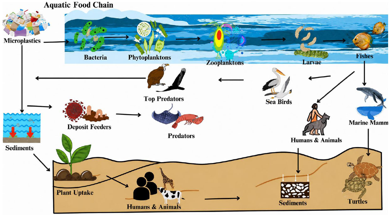
In the atmosphere, MNPs are transported, accumulated, and dispersed through both natural processes and anthropogenic activities. Physical and biological transport comprises wind erosion, soil erosion, water runoff, bioturbation by soil fauna, biofouling, trophic transfer, and biological fragmentation. Anthropogenic activities comprise construction dust, tire abrasion, personal care products, textile fibers, sewage sludge and compost, laundry, agricultural practices, resuspension of contaminated soils, etc. (Fig. 1). These activities allow accumulation and distribution of MNPs in terrestrial, aquatic, and atmospheric regions (Bergmann et al., 2017; Allen et al., 2019).
2.2. Human Exposure Pathways to Micro-and Nanoplastics
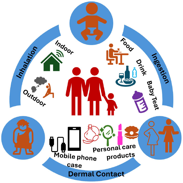
Humans are the primary contributors to the dissemination of MNPs into the environment through ingestion, dermal contact, and inhalation (Fig. 2). A comprehensive assessment of these direct and indirect exposure routes is essential for accurately evaluating the health risks posed by MNPs.
2.2.1. Inhalation
Inhalation is the primary factor driving the translocation of MNPs in the environment and within organisms. It results in various respiratory and systemic disorders. It facilitates exposure to MNPs through both indoor and outdoor air.
2.2.1.1. Indoor Air
Since individuals spend almost 89% of their time indoors, indoor air is a major source of airborne MNP exposure. Reported concentrations of MNPs indoors vary widely depending on room functions, human activities, and ventilation. For example, 1.1-18.2 fibers/m3 in Paris apartments, 4.0-59.4 fibers/m3 in offices, and even higher levels in nail salons/e-waste facilities were reported (Dris et al., 2017; Chen et al., 2022). Deposition rates of MNPs in indoor environments also differ significantly, with homes exhibiting the highest accumulation. Most indoor MNPs are fibers larger than 20 µm, though smaller MNPs (< 2.5 µm) are of greater concern due to their ability to remain airborne and reach deeper lung regions. Infants, children, and older adults experience higher exposure because they spend more time indoors and frequently come into contact with dust, which can contain high concentrations of MNPs (Zhang et al., 2020; Nematollahi et al., 2022). Thus, indoor activity patterns, environment, and age strongly influence inhalation risk (Fig. 2).
2.2.1.2. Outdoor Air
Outdoor MNP concentrations are generally lower due to greater air movement; however, clear indoor-outdoor and urban-rural contrasts exist. In Wenzhou, China, indoor MNP concentrations (1,583 ± 1,180 particles/m3) exceed outdoor levels (189 ± 85 particles/m3) by an order of magnitude, and similar patterns are observed in European cities (Dris et al., 2017; Liao et al., 2021). Urban regions typically show higher levels than rural areas, and estimated daily inhalation varies regionally, from nearly 21 particles/day in Shanghai to several thousand particles/day in some parts of Wenzhou (Liao et al., 2021; Liu et al., 2019). Although outdoor exposure is usually lower than indoor exposure, microenvironments such as roadsides, construction zones, and landfills may exhibit elevated concentrations that warrant further study (Cox et al., 2019; Zhang et al., 2020).
2.2.2. Ingestion
Ingestion is also the main factor driving the translocation and accumulation of MNPs in organisms and the environment (Fig. 2). Organisms ingest various food items that are contaminated with MNPs. Then these MNPs are transported through food webs and enter the gut. There, these MNPs disrupt the integrity of the gut epithelium and result in various gastrointestinal disorders.
2.2.2.1. Seafood Ingestion
Seafood intake is a notable dietary route for human exposure to MNPs. Numerous marine organisms, such as cephalopods, crustaceans, bivalves, and fish, ingest MNPs from contaminated seawater and deposited sediments due to their resemblance to various natural foods (Jâms et al., 2020; Curren et al., 2020). Because they are consumed whole by humans, oysters and mussels are more responsible for translocation of MNPs through food webs. In addition, MNPs are carriers of numerous chemicals, heavy metals, organic pollutants, and pathogens that cause oxidative stress and inflammation in human beings (Wootton et al., 2021). Oysters from polluted areas have been reported to contain nearly 100 particles per individual. (Expósito et al., 2022). Moreover, marketed samples of mollusks generally contain fewer MNPs than directly caught samples due to cleaning (Nicole, 2021). Crustaceans accumulate MNPs primarily in inedible organs, where levels are about four times higher than in edible tissues (Yin et al., 2022). Interestingly, avoiding the consumption of the intestinal parts of shrimp and crab can reduce exposure. Accurate intake estimates should focus on MPs in edible portions.
2.2.2.2. Drinking Water and Beverages Ingestion
Bottled water contains substantially more MNPs than tap water (Oßmann et al., 2018; Akhbarizadeh et al., 2020). Some glass-bottled beverages were reported to contain more than 6,000 particles/L (Oßmann et al., 2018). The size of most particles was 1-5 μm. Tap water generally shows lower levels (up to nearly 930 particles/L), and boiling does not remove MNPs. Annual adult intake is estimated at ~ 4.6 × 10⁵ particles from tap water and ~3.6 × 10⁷ from bottled water.
MNPs are also present in beverages such as tea, milk, and beer, largely due to contaminated water (Shruti et al., 2020; da Costa Filho et al., 2021). Beer shows variable contamination depending on the analytical method used. Tea bags release MNPs from a few to thousands of particles, depending on the region (Ali et al., 2023). Ongoing monitoring and standards, especially for bottled water, are needed to manage exposure.
2.2.2.3. Honey, Salt, and Sugar Ingestion
MNPs have also been detected in multiple food additives, such as sugar, honey, and salt (Ali et al., 2023; Peixoto et al., 2019). The shape of most MNPs was fibrous and fragmented (Peixoto et al., 2019). The annual consumption of MNPs by adults through salt was estimated at 35.8-36,172 particles (Lee et al., 2021). Sugar samples from Bangladesh exhibited high contamination (nearly 344 particles/g), while honey generally contained lower levels (22-114 particles/L (Diaz-Basantes et al., 2020). Other common seasonings and condiments, such as cooking oil, monosodium glutamate, and soy sauce, also warrant investigation to fully assess dietary MNP exposure (Fig. 2).
2.2.2.4. Crops and Livestock Ingestion
Field data on MNP contamination in crops and livestock are limited. Laboratory studies show that numerous plants, including carrots, lettuce, rice, wheat, and peanuts, can take up and accumulate MNPs in edible tissues (Li et al., 2020; Dong et al., 2021). While natural uptake is generally low, long-term consumption of contaminated crops may pose health risks. Livestock can also accumulate MPs. For example, nearly 46 MNPs were reported per chicken gizzard (Huerta Lwanga et al., 2017). MNPs have also been detected in edible snails. Food items can also acquire MNPs during production, processing, packaging, and transport (Fig. 2). Rice samples from Australia contained an average of 67 ± 26 µg/g MNPs, and packaged meats showed 4-18.7 particles/kg (Dessì et al., 2021; Kedzierski et al., 2020). Dust ingestion also contributes to intake of MNPs. Adults consume nearly 4.0 × 10²-2.5 × 10⁴ particles annually, and infants even more. However, data remain scarce for major foods such as fruits, vegetables, meat, and grains. Therefore, accurate exposure assessments must consider local dietary patterns and measure MNP concentrations in staple foods (Danopoulos et al., 2020; Domenech et al., 2021; Zhu et al., 2022).
2.2.3. Dermal Contact
Human skin is routinely exposed to MNPs through personal care products (Fig. 2), such as toothpaste, sunscreens, soaps, and face washes (Rahman et al., 2021; Lett et al., 2021). Before regulatory restrictions in 2018, microplastics were widely used as exfoliants, raising concerns that nanoparticles could penetrate the skin barrier (Fendall and Sewell, 2009; Sykes et al., 2014). Additional dermal exposure may occur from wear debris generated by plastic components in prostheses and from airborne MNPs settling onto the skin (Wright et al., 2017; Abafe et al., 2023). Although no quantitative estimates exist for dermal absorption, the potential risks, especially from compromised skin exposed to nanoplastics, should not be overlooked.
3. PHYSICOCHEMICAL PROPERTIES OF MICRO- AND NANOPLASTICS
MNPs exhibit heterogeneous physicochemical characteristics that influence their transport, environmental fate, and biological consequences. Nanoplastics have higher colloidal stability and a larger surface area, which increase their reactivity and capacity to penetrate biological barriers (Xia et al., 2026; Andrady, 2017). Morphological variations include fragments, fibers, films, and spheres. Particle shape influences bioavailability, aggregation behavior, and uptake by organisms, where fibrous shapes demonstrate greater respiratory and gastrointestinal retention (Cole et al., 2011). Environmental weathering leads to the evolution of surface chemistry, with oxidation and chain scission followed by the formation of reactive functional groups that alter hydrophobicity, surface charges, and protein and pollutant interactions (Gewert et al., 2015; Lamber and Wagner, 2016). Crystallinity, density, glass transition temperature, and additive content are determined by the polymer type (e.g., PP, PS, PE, PVC, PET), thereby affecting environmental persistence and the leaching of plasticizers, stabilizers, and flame retardants (Rochman et al., 2011). The density variations guide the mode of sedimentation, floating, or suspension of the particle, impacting the exposure route and trophic transfer (Hidalgo-Ruz et al., 2012). In addition, mechanical resilience and degradation rates are determined by molecular weight and crystallinity. Low-crystallinity polymers exhibit higher degradation rates under UV and oxidative conditions. (Andrady 2011). These physicochemical features regulate the sorption of organic contaminants and metals, alter interactions with biological membranes, and determine the accumulation of MNPs in ecosystems and human tissues (Teuten et al., 2009; Schwabl et al., 2019).
The environmental behavior and biological impacts of MNPs are strongly governed by their physicochemical properties, leading to a complex structure-activity relationship. Particle size is a primary determinant of mobility and biological interactions. Due to high surface-to-volume ratios, high colloidal stability, and enhanced ability to penetrate biological barriers, NPs nanoplastics facilitate cellular uptake and systemic distribution. Surface charge and functional groups regulate interactions with natural organic matter, proteins, and cellular membranes. This, in turn, influences aggregation behavior, environmental persistence, and uptake pathways. The morphology of MNPs, such as spherical fragments, fibrous structures, and irregular particles, affects transport, sedimentation rates, and ingestion probability in aquatic and terrestrial organisms. Polymer composition further determines density, hydrophobicity, additive release, and sorption affinity for environmental pollutants. Heavy metals and persistent organic pollutants may further amplify indirect toxicity through vector effects. In addition, environmental aging processes such as ultraviolet irradiation, mechanical abrasion, and oxidative weathering progressively change the surface chemistry and roughness of MNPs. This leads to increased reactivity, pollutant adsorption capacity, and interactions with microbial biofilms.
4. TOXICOLOGICAL EFFECTS OF MICRO- AND NANOPLASTICS IN THE ECOSYSTEM
MNPs exert toxic effects across terrestrial and aquatic ecosystems by disrupting biochemical, physiological, and cellular processes in exposed organisms. Their small size, persistence, and surface reactivity enable bioaccumulation, trophic transfer, and interactions with biological barriers, leading to inflammation, oxidative stress, metabolic dysfunction, reproductive impairment, reduced growth, and decreased survival.
4.1. Cellular, Physiological and Toxicological Consequences of Micro- and Nanoplastics Exposure In Terrestrial Vertebrates
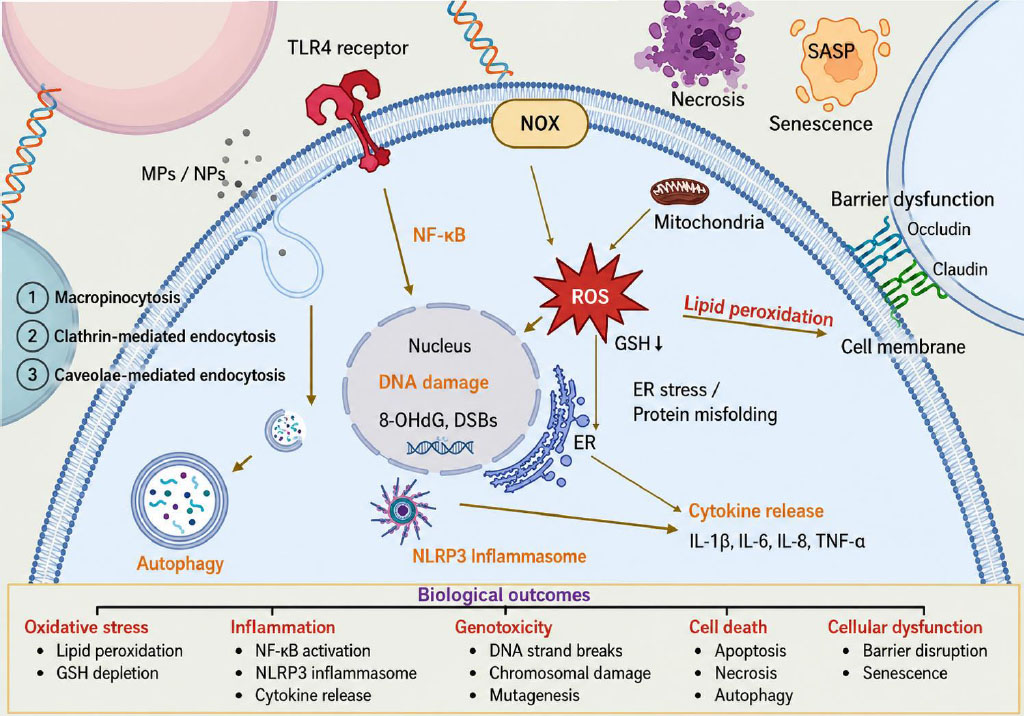
Exposure of vertebrates to MNPs results in direct interactions between MNPs and cellular components and various physiological systems. At the cellular level, MNPs interact with cell membranes and intracellular components like the nucleus, cytoplasm, mitochondria, endoplasmic reticulum, lysosome, Golgi apparatus, etc. (Haldar et al., 2023; Bai et al., 2024; Tripathi and Goshisht, 2022). This interaction leads to the initiation of a cascade of toxicity responses. Due to their small size, nanoplastics generally penetrate cellular membranes via active or passive diffusion, thereby generating Reactive Oxygen Species (ROS). The generated ROS trigger oxidative stress, inflammation, lipid peroxidation, mitochondrial disruption, DNA damage, protein oxidation, and disruption of cellular homeostasis (Guan et al., 2023; Amereh et al., 2020; Lin et al., 2025). Chronic exposure of organisms to MNPs leads to multisystem physiological disturbances at the organismal level. MNPs accumulate in several organs, such as the lungs, gastrointestinal tract, kidneys, liver, spleen, and heart, and lead to disruption of epithelial barriers (Liu et al., 2023) [7].
Microplastics are primarily taken up by phagocytosis, whereas nanoplastics enter more rapidly via endocytosis, macropinocytosis, and direct membrane penetration. Once inside, MNPs cross the gut lining, pass through the air-blood barrier following inhalation, and subsequently enter the lymphatic system, thereby contributing to widespread distribution, as shown in Fig. (3)(Lin et al., 2025). Many particles persist in tissues because they resist degradation and form a protein corona that alters how cells recognize them. Evidence shows that MNPs accumulate in diverse organs. (Momeni et al., 2025). These processes suggest that chronic, low-dose exposure may lead to long-term health risks that require careful evaluation. This section summarizes recent evidence of MNPs toxicity in terrestrial vertebrates and investigates how cellular mechanisms shape the vulnerability of different physiological systems (Tables 1 and 2, and Fig. 3).
| Human Anatomical System | Human Sample | Micro- and Nanoplastics | Toxicity profile | Ref. | |||
|---|---|---|---|---|---|---|---|
| Type | Size | Dose | Exposure Duration | ||||
| Respiratory | Pulmonary alveolar cells (HPAEpIC) |
PSNPs | 40 nm | - | 24 h | Increased cell viability and MMP-9; surfactant protein A dysregulation. |
(Yang et al,. 2021) |
| Alveolar epithelial cells (A549) |
PSNPs | 25 and 70 nm | 1.14-25 μg/mL | 24-48 h | Decreased cell viability and increased apoptosis; increased TNF-α, IL-8, and NF-κB. | (Xu et al., 2019) | |
| Digestive | Colon adenocarcinoma cells (Caco-2, HT29) |
PE-MPs | 1-10 μm |
21mg/8 mL | 14 days | Increased pathobionts; decreased beneficial bacteria (e.g., Akkermansia); intestinal homeostasis loss. | (Fournier et al., 2023) |
| Cardiovascular | Embryonic stem cells (hiPSCs) |
PSNPs | 40 nm | 1 × 10⁹/mL | 24 h | Reduced heart valve development; decreased LEFTY1/2; increased CA4, OCLM. |
(Bojic et al., 2020) |
| Human Umbilical Vein Endothelial Cells (HUVECs) |
PSNPs | 100 nm and 500 nm | 5-100 μg/mL | - | Increased LDH release, membrane damage, and autophagy; ER stress. |
(Lu et al., 2022) | |
| Reproductive | Testicular cells (NTE) |
PSNPs | 50 nm | 200 μg/mL | 24 h | Increased JNK1/2/3, TNF-α; Dysregulated PI3K-AKT Pathway; oxidative stress. |
(Xiao et al., 2023) |
| Nervous | Forebrain cortical spheroids (3D model) |
PSMPs | 1 μm and 10 μm |
5-100 μg/mL | 4-30 days | Decreased cortical layer VI markers; decreased neuronal maturation; altered Nestin expression. | Hua et al., 2022 |
| Targeted Aspect/System | Nanoplastic | Toxicity Profile | Ref. | ||||
|---|---|---|---|---|---|---|---|
| Type | Size | Exposure Route | Dose | Exposure Duration | |||
| PS | ~50 nm | Oral | 0-10 mg/kg | 30 days | Modified gut microbiota and mucus-related genes; no inflammation in major organs. | (Xiao et al., 2022) | |
| PS | ~80 nm and 500 nm |
Oral | 5-50 mg/kg | 2 weeks | Accumulated in liver < kidneys < intestine; increased COX-2, iNOS, cytokines, ROS, superoxide dismutase activity, and Nrf2 expression. | (Choi et al., 2021) | |
| PS | 50 nm | Drinking water | 0.1-10 mg/L | 32 weeks | Intestinal barrier damage (decreased tight junctions); oxidative stress, villus erosion, immune disruption (increased B cells, decreased CD8+ T cells). | (Li et al., 2024) | |
| PS | ~100 nm | Oral | 1 mg/day | 28 days | Crohn’s-like ileitis; intestinal epithelial Necroptosis (RIPK3/MLKL); impaired mitophagy (lysosomal dysfunction). | (Huang et al., 2022) | |
| Brain and behavior |
PS | 50 nm | Oral | 0.5-50 mg/kg | 7 days | Crossed blood-brain barrier; neuronal damage; microglial activation; increased ROS, TNF-α release, NF-κB activation, necroptosis. | (Shan et al., 2022) |
| PS | 50 nm | Oral | 0.25-250 mg/kg | 28 days | Parkinson’s-like neurodegeneration, mitochondrial dysfunction; astrocyte inflammation, excitatory neurons most vulnerable. | (Liang et al., 2022) | |
| PS | 25 nm | Oral | 0-50 mg/kg | 6 months | Cognitive decline; increased ROS; DNA damage; synaptic dysfunction, altered expression of mRNAs, miRNAs, circRNAs. | (Chu et al., 2022) | |
| PS | 80 nm | Inhalation (aerosol) | - | - | Crossed brain tissue; suppressed acetylcholinesterase activity; decreased locomotor activity. | (Liu et al., 2022) | |
| Reproductive system |
PS | 500 nm | Oral | 0-1.5 mg/day | 90 days | Damaged seminiferous tubules; impaired BTB integrity via MAPK/Nrf2 suppression; sperm Apoptosis. |
(Li et al., 2021) |
| PS | ~39 nm | Oral | 1-10 mg/kg/day | 5 weeks | Decreased luteinizing hormone, testosterone, follicle-stimulating hormone; abnormal sperm; sperm DNA damage; disrupted hypothalamic-pituitary axis. | (Amereh et al., 2022) | |
| PS | 50 nm | Oral | 0.2 mg/mL | 60 days | Leydig cell hyperplasia; Lower testis weight; poor sperm quality; vascular congestion; structural damage. | (Sharma et al., 2024) | |
| Systemic | PET | ~96 nm | Oral | 200 mg/kg | 30 days | Kidney damage (renal corpuscles/tubules degeneration); increased blood urea nitrogen, malondialdehyde, creatinine; decreased glutathione. | (Kamel et al., 2024) |
| PS | 40 and 200 nm | Oral | 0.01-0.1 mg/day | -- | Accumulated in organs; male testis toxicity (reduced testosterone, abnormal sperm); anxiety-like behavior; immune disruption. |
(Nikolic et al., 2022) | |
| PS | 25-50 nm | Oral | 1-10 mg/kg/day |
5 weeks | Escalated ROS; altered liver and kidney markers; stress-related biochemical changes; disrupted energy metabolism. | (Babaei et al., 2022) | |
| Transgenerational | PS | 100 nm | Maternal | 0.1-10 mg/L | - | Impaired testis development and sperm count; reduced offspring body weight; male offspring liver damage, oxidative stress. | (Huang et al., 2022) |
4.2. Toxicity Of Micro- And Nanoplastics In Terrestrial Invertebrates
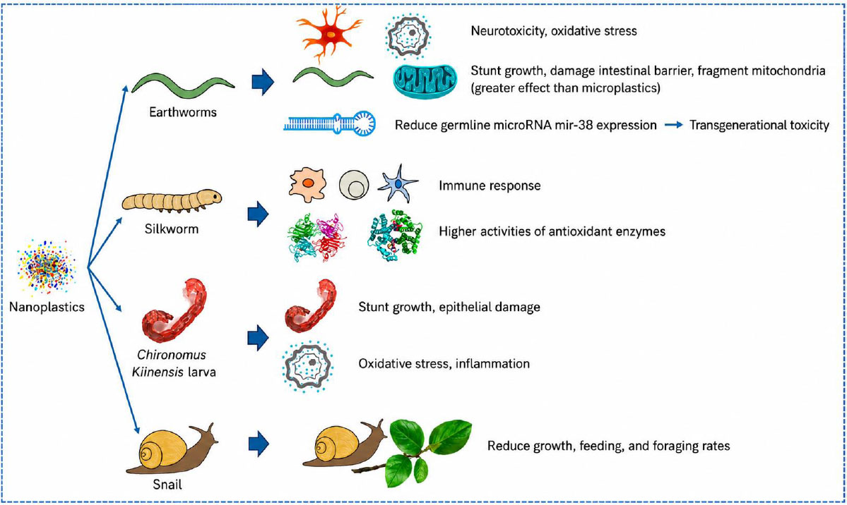
Terrestrial invertebrate studies on nanoplastic toxicity focus primarily on earthworms and Caenorhabditis elegans (C. elegans). Exposure of C. elegans to 20-25 nm polystyrene nanoplastics, even at low concentrations, impaired intestinal integrity, growth, and movement (Liu et al., 2021). The nanoplastics accumulate in the gut and spread to other tissues, resulting in mitochondrial damage (Fig. 4). 20 nm nanoplastics produced stronger and longer-lasting transgenerational toxicity than 100 nm particles. Additionally, 20 nm nanoplastics reduced germline microRNA mir-38, and its downregulation drove multigenerational toxicity via the NDK-1-KSR-1/2 signaling pathway (Hua et al., 2022). Exposure of insects to polystyrene nanoplastics suppressed immune gene expression and antioxidant activity in Bombyx mori I (Muhammad et al., 2021). This suppression led to higher mortality following bacterial infection. The exposure of Chironomus kiinensis larvae to amino-functionalized positively charged polystyrene (PS-NH2) nanoplastics caused greater gut accumulation, oxidative stress, microbiota disruption, and epithelial injury as compared to negatively charged particles (Zhang et al., 2026). A terrestrial food chain study reported that nanoplastics accumulated in mung beans grown in contaminated soil when transferred to snails; the nanoplastics reduced growth, feeding, and gut health of snails. This study demonstrated trophic transfer and biological impacts across terrestrial systems (Li et al., 2026). The effects of nanoplastics on various terrestrial invertebrates have been presented in Fig. (4) (Tang, 2025).
4.3. Toxicological Effects of Micro- and Nanoplastics in Aquatic Organisms
Nanoplastics pose significant toxicological threats to aquatic animals such as fishes, crustaceans, and bivalves (Table 3) due to their high surface reactivity, small size, and ability to penetrate diverse biological barriers (Contino et al., 2023; Sun et al., 2024). In crustaceans, nanoplastics disrupt gut function, alter immune responses, induce oxidative stress, impair molting and growth, and trigger apoptosis (Machado et al., 2021). Bivalves, which continuously filter large volumes of water, readily accumulate nanoplastics in their gills and digestive glands, leading to genotoxicity, lysosomal instability, oxidative and inflammatory damage, and compromised immune and reproductive functions (Liu et al., 2021). In fish, MNPs accumulate in multiple organelles and cause inflammation, oxidative stress, and genotoxicity (Sun et al., 2024).
| Species/Target Organism | Nanoplastic Type | Size | Concentration | Duration | Toxicity | Ref |
|---|---|---|---|---|---|---|
|
Zebrafish (D. rerio) |
Polystyrene | ~70 nm | 0.5 and 1.5 ppm | 7 days and 7 weeks |
Accumulation in brain, liver, intestine, and gonads; disruption of lipid/energy metabolism; oxidative stress and behavioral alterations (aggression, locomotion, and shoaling, circadian rhythm). | (Sarasamma et al., 2020) |
|
Fathead minnow (P. promelas) |
Polystyrene | 50 nm | 5 μg/L (injection, ingestion) |
48 h | Downregulation of immune genes (mst1, ncf, c3); immunotoxicity in liver and kidney. | (Elizalde-Velazquez et al., 2020) |
|
Tilapia (O. niloticus) |
Polystyrene | 100 nm | 20 mg/L | 7 days + 7day recovery |
Persistent transcriptomic and metabolic toxicity. | (Pang et al., 2021) |
|
Largemouth bass (M. salmoides) |
Polystyrene | 100 nm | 10 and 100 μg/L | 7-9 days | Alterations in gill, liver, and intestine; transient oxidative stress. | (Chen et al., 2022) |
|
Common carp (C. carpio) |
Polystyrene | 50-400 nm | 1000 μg/L | 28 days | Myocardial inflammation and apoptosis; increased TLR4, NOX2, reactive oxygen species, malondialdehyde; ↓ antioxidant enzymes; Th1 immune shift. | (Wu et al., 2022) |
| Mussel (M. galloprovincialis) | Polystyrene nanoplastics | (100 nm) | 0.5 and 5 mg・L−1 |
7 days | Disruption of ion regulation, oxidative balance, and energy metabolism in gills. | (Wang et al., 2023) |
|
Mediterranean mussel (M. galloprovincialis |
Amino-modified polystyrene nanoplastics |
(50 nm) | 10 μg・L−1, 24 h × 2 |
(72 h interval) | Mitochondrial and lysosomal stress, altered gene expression, reduced lysozyme; re-exposure restored the functions and enhanced immune gene expression. | (Auguste et al., 2020) |
|
Manila clam (R. philippinarum) |
Pd-doped polystyrene nanoplastics |
(139.5 nm, +43.8 mV) |
0.02 and 2 mg・L−1 |
14 days | High dose induced ROS, lipid peroxidation, tissue damage and apoptosis; altered gut microbiota at low dose. | (Zhou et al., 2023) |
4.4. Toxicological Effects of Micro- and Nanoplastics on Plant Health
Interaction between MNPs and plants may occur through various routes, including atmospheric, soil, and aquatic environments (Chen et al., 2025). Leaf cuticles regulate water loss and gas exchange. This serves as the main defense against micro- and nanoplastics. However, MNPs smaller than 5 nm can easily overcome this defense through hydrophilic and lipophilic routes (da Costa et al., 2016; Wang et al., 2023). Moreover, stomata, being 3-10 µm wide, serve as primary entry points for MNPs. After internalization through stomata, MNPs are unidirectionally translocated to different plant tissues via the vascular system (Du et al., 2025). Afterwards, MNPs induce cellular, physiological, and organ-level toxicity (Canniff and Hoang, 2018; Jia et al., 2023). Accumulation of MNPs triggers oxidative stress, reduced mitosis, membrane damage, disruption of nutrient transport, reduced metabolic enzyme activity, and ultimately gene expression in various species such as Triticum aestivum, Phaseolus vulgaris, Hordeum vulgare, Arabidopsis thaliana, etc. (Ranjan et al., 2021; Colzi et al., 2022; Hernández-Sánchez et al., 2025). Consequently, length, activity, and biomass of different plant parts such as roots and shoots are inhibited.
MNPs also affect photosynthesis in plants. However, these effects depend upon concentration, size, and environmental conditions (Tan et al., 2025). They lead to the production of ROS, impaired electron transport, disrupted chlorophyll synthesis, reduced photosynthetic pigments, reduced photosystem efficiency, and altered resource uptake/carbon fixation (Roy et al., 2024). Ultimately, MNPs cause ROS production, membrane lipid peroxidation, disruption of cellular integrity, reduced efficiency of the defense system/nutrient uptake/metabolism, and altered gene expression. In addition, heavy metals, pathogens, and organic pollutants carried by MNPs further affect photosynthetic efficiency, plant defense, and pathogen persistence and dissemination (Teuten et al., 2009; Shah et al., 2023). This section explores the toxicity of micro- and nanoplastics across various plant species and summarizes the findings in Table 4.
| Parameter | Plant Species | MNPs Type | MNPs Size/Shape | Exposure Duration | Effects on Aquatic Environment | Effects in Soil Environment | Mechanism of Action | Ref. |
|---|---|---|---|---|---|---|---|---|
| Root Morphology |
Lactuca sativa, Allium cepa |
PP, PE, PS |
Fibers and spheres | 14-28 days | Increased lateral root formation in some aquatic species due to nutrient redistribution. | Inhibited root elongation and reduced root hair density; soil compaction and abrasion effects. | Physical obstruction; nutrient leaching and competition. | (Giorgetti et al., 2020; Li et al., 2020) |
| Germination Rate |
Lactuca sativa and Triticum aestivum |
PVC, PE, PS |
Fragments and fibers (< 5 mm) |
7-21 days | Generally unaffected or slightly enhanced (duckweed) owing to constant hydration and MNPs adsorption. | Decreased germination in crop plants (e.g., lettuce, wheat, etc.) due to impaired water and nutrient uptake. | MNPs obstruct seed pores and adsorb allelochemicals | (Qi et al., 2018; Lian et al., 2021) |
| Chlorophyll Content | Spirodela polyrhiza and Lemna minor | PS, PLA, PET |
Fibers, Nanospheres (<100 nm) |
7-21 days | Varying effects: stable or slightly elevated in duckweed; reduced in submerged macrophytes. | Reduction in chlorophyll a and b owing to oxidative stress and inhibited nutrient uptake. | ROS formation; nutrient chelation and enhancement | (Mateos-Cardenas et al., 2019) |
| Nutrient Uptake |
Lactuca sativa, Zea mays |
PS, PE |
Particles | 7-30 days | Mixed outcomes: sometimes enhanced uptake as MNPs act as nutrient carriers. | Reduced uptake of P, N, Mg, and K owing to root disruption and ion sorption by MNPs. | Physicochemical interactions of MNPs with nutrient ions | (Gao et al., 2019; Sun et al., 2021) |
| Gene Expression | Oryza sativa, Arabidopsis thaliana | PS, PE | Nanoplastics, Fragments | 7-14 days | Upregulation of stress- linked genes (e.g., glutathione metabolism and heat-shock proteins). |
Downregulation of genes controlling growth (e.g., aquaporins, auxin signaling, etc.). | MNPs-induced modulation of hormonal and stress pathways | (Taylor et al., 2020; Wu et al., 2022) |
| Biomass Accumulation |
Oryza sativa, Triticum aestivum | PVC, PE | Spheres, fragments (<1 mm) |
14-30 days | Neutral or increased biomass owing to improved nutrient availability through MNPs surface sorption. | Decrease in root and shoot biomass (10-30%) owing to stress and reduced photosynthetic efficiency. | Disrupted root-soil interaction versus enhanced nutrient absorption | (Taylor et al., 2020; Zhang et al., 2021) |
| Reproductive Development |
Solanum lycopersicum, Arabidopsis thaliana, |
PS, PE | Fragments | >30 days | Limited data: most studies focus on early growth stages in aquatic species. | Reduced flowering, pollen viability, and seed production. | Stress-driven reallocation of energy from reproduction to defense. | (Lian et al., 2021; Nei et al., 2024) (Lian et al., 2021; nei et al., 2024) [126,133]. |
| Oxidative Stress (ROS) |
Lemna minor, Lactuca sativa | PE, PS | Nano/microspheres | 10-21 days | Increased lipid peroxidation, ROS, and antioxidant defense. | Elevated ROS, lipid peroxidation, and increased antioxidant enzymes (SOD, CAT, POD, etc.). | Cellular injury from ROS accumulation. | (Mateos-Cardenas et al., 2019; Gao et al., 2021). |
| Photosynthetic Efficiency |
Lactuca sativa, Lemna minor |
PE, PS, PVC |
Fibers, particles | 7-28 days | Generally stable or improved performance in floating plants (e.g., duckweed). | Lower photosynthetic rate, stomatal conductance, and transpiration. | Stomatal blockage in terrestrial plants vs. buoyancy advantage in aquatic species. | (Mateos-Cardenas et al., 2019; Gao et al., 2021). |
The current understanding of the toxicological effects of MNPs is derived from multiple levels of evidence, including in vitro cellular studies, in vivo animal experiments, and emerging human exposure data (Araújo et al., 2025; Thomas et al., 2026). However, each level of evidence has specific strengths and limitations when extrapolating experimental findings to real-world human exposure scenarios. A summary of these evidence levels and their associated limitations is presented in Table 5.
| Evidence Level | Study Type | Typical Experimental Model | Key Insights Provided | Major Limitations for Real-World Risk Assessment |
|---|---|---|---|---|
| in vitro studies | Cell culture experiments. | Human or animal cell lines (e.g., epithelial cells, macrophages, intestinal cells). | Provide mechanistic insights such as oxidative stress, cytotoxicity, inflammation, and cellular uptake of MNPs. | Simplified biological systems; lack of whole-organism interactions; exposure conditions often not environmentally realistic. |
| in vivo animal studies | Controlled laboratory experiments. | Rodents, zebrafish, fish, and other vertebrates. | Evaluate bioaccumulation, organ distribution, physiological responses, and systemic toxicity. | Species-specific physiological differences may limit direct extrapolation to humans; exposure doses often exceed environmental levels. |
| Ex vivo studies | Tissue-based experimental systems. | Isolated tissues or organ cultures. | Provide insights into tissue-level responses and biological interactions with MNPs. | Limited representation of systemic biological processes. |
| Human observational evidence | Environmental or biomonitoring studies. | Detection of microplastics in human tissues, blood, placenta, lungs, or feces. | Direct evidence of human exposure to microplastics and potential bioaccumulation. | Limited data on health outcomes; causality between exposure and disease remains unclear. |
| Human health risk extrapolation | Risk modeling and comparative toxicology. | Integration of laboratory data with environmental exposure estimates. | Helps estimate potential human health risks and exposure thresholds. | High uncertainty due to limited exposure data, variability in particle characteristics, and lack of long-term epidemiological studies. |
Recent studies have suggested that nanoplastics may enter plant tissues through several potential pathways, including stomatal openings, cuticular diffusion, and root uptake followed by vascular transport (Vleeming et al., 2026; Zhu et al., 2026). However, the relative importance of these pathways remains uncertain and appears to be highly context-dependent. Evidence for stomatal entry has largely been derived from controlled experimental studies, where nanoscale particles were shown to penetrate leaf surfaces under specific exposure conditions. Similarly, the diffusion of nanoplastics through the cuticular layer has been proposed as a possible mechanism, particularly for extremely small particles with appropriate surface properties (Bhati et al., 2026). Nevertheless, these transport processes cannot yet be considered universal mechanisms for nanoplastic uptake in plants. Their occurrence and efficiency may depend on several factors, including particle size, surface functionalization, plant species, leaf morphology, environmental conditions, and exposure concentrations (Piovesan et al., 2026). Moreover, many existing studies rely on laboratory-based exposure systems that may not fully represent realistic environmental scenarios. Therefore, further research is required to clarify the dominant uptake pathways and transport dynamics of nanoplastics in plants under environmentally relevant conditions.
4.5. Micro- and Nanoplastics as Vectors of Co-Contaminants: Combined toxicity and Food-Chain Transfer
MNPs are increasingly recognized not only as emerging pollutants themselves but also as effective vectors for a wide range of environmental contaminants like persistent organic pollutants (POPs), heavy metals, pesticides, pharmaceuticals, and pathogenic microorganisms (Li et al., 2025). Due to their large surface area, hydrophobicity, and diverse surface functional groups, MNPs can readily adsorb and concentrate chemical and biological pollutants from surrounding environmental matrices such as water, sediments, soil, and atmospheric particles. This capacity to bind multiple contaminants significantly enhances the complexity of MNP-associated environmental risks and represents a major research hotspot in the field of environmental toxicology.
4.5.1. Adsorption and Interaction Mechanisms of Co-Contaminants
The adsorption of pollutants onto MNP surfaces occurs through several physicochemical mechanisms, including hydrophobic interactions, electrostatic attraction, van der Waals forces, π-π interactions, and surface complexation (Rai et al., 2022). Environmental aging processes such as UV irradiation, mechanical abrasion, and oxidative weathering can further modify the surface chemistry of plastics by introducing oxygen-containing functional groups (e.g., hydroxyl and carbonyl groups), thereby enhancing their adsorption capacity for metal ions and organic molecules (Sekar et al., 2026). Consequently, aged MNPs often exhibit significantly higher affinities for contaminants compared with pristine plastics.
Heavy metals such as lead (Pb), cadmium (Cd), chromium (Cr), copper (Cu), and zinc (Zn) have been shown to strongly associate with MNP surfaces through electrostatic interactions and complexation with surface functional groups (Mohamed et al., 2025). Similarly, hydrophobic organic pollutants, including polycyclic aromatic hydrocarbons (PAHs), polychlorinated biphenyls (PCBs), pesticides, and pharmaceutical residues, can partition onto plastic surfaces through hydrophobic and π-π interactions. These interactions enable MNPs to act as mobile carriers of environmental pollutants, facilitating their long-distance transport and redistribution across ecosystems.
4.5.2. Combined Toxic Effects of MNPs and Associated Pollutants
The co-occurrence of MNPs with other contaminants may lead to additive, synergistic, or antagonistic toxic effects in biological systems. When organisms ingest MNPs carrying adsorbed pollutants, the particles can serve as delivery vehicles, increasing the bioavailability of these contaminants and promoting their accumulation in tissues (Samaei et al., 2025). Several studies have demonstrated that co-exposure to MNPs and heavy metals can exacerbate oxidative stress, inflammatory responses, mitochondrial dysfunction, and cellular damage (Sun et al., 2025; Devi et al., 2025). Similarly, MNPs interacting with organic pollutants can amplify toxicological responses by enhancing membrane permeability, endocrine disruption, and metabolic disturbances. For instance, the combined exposure of aquatic organisms to MNPs and hydrophobic organic contaminants has been associated with elevated ROS production, DNA damage, and impaired physiological functions.
In addition to chemical pollutants, MNPs can also act as substrates for microbial colonization, forming complex microbial communities known as the “plastisphere.” These biofilms may include pathogenic bacteria, antibiotic-resistant microbes, and harmful algae, potentially facilitating the dissemination of pathogens and antibiotic resistance genes in aquatic and terrestrial environments. The coexistence of MNPs, toxic chemicals, and microorganisms therefore creates a multifactorial exposure scenario that may significantly increase ecological and human health risks.
Chu et al. 2024 studied the effects of polystyrene microplastics (1 μm) and nanoplastics (100 nm) on growth, oxidative stress, tissue damage, metabolism, and gut microbiota of rare minnows, Gobiocypris rarus. Chemical and multi-omics studies were employed to evaluate the results. Both particle types inhibited growth and development, induced histopathological lesions, and increased oxidative stress, with the effects changing with size and concentration. High microplastic exposure led to considerable shifts in the gut microbiota, while high nanoplastic exposure reduced diversity. Ding et al. 2020 studied the effects of polystyrene nano- and microplastics (0.3, 5, and 70-90 μm, at 95, 92, and 112 μg/L) on red tilapia. After 14 days, larger particles (70-90 μm) accumulated the most. Microplastics (5 and 70-90 μm) caused more oxidative damage than nanoplastics. Polystyrene nanoplastics significantly reduced CYP3A-related enzymes, but liver CYP enzymes were less affected by microplastics. Microplastics as small as 5 μm were shown to dramatically impair acetylcholinesterase activity in the brain. Metabolomics found changed metabolites for 0.3, 5, and 70-90 μm doses, with a similar impact on tyrosine metabolism. Overall, μm-sized particles induced more physiological stress than nanoplastics.
Chen et al. (2017) investigated how MPs microplastics (45 μm) and NPs nanoplastics (50 nm), both directly and indirectly, affect the locomotion of zebrafish larvae. The larvae were exposed to 1 mg/L of MPs microplastics and NPs nanoplastics for 48 and 72 h. Their findings showed that microplastics had minimal impact, apart from an increase in the expression of the visual gene zfrho. In contrast, nanoplastics caused a 22% reduction in larval movement during the final dark phase, suppressed acetylcholinesterase activity by 40%, reduced body length by 6%, and significantly increased the expression of gfap, α1-tubulin, zfrho, and zfblue genes. Latchere et al. 2023 compared the toxicity of polystyrene nanoplastics (200 nm) and environmentally derived microplastics (1.2 to 300 μm) and nanoplastics (235 nm) on the freshwater bivalve Corbicula fluminea (Muller, 1774). They exposed the organisms to 0.008, 10, or 100 μgL−1 for 21 days, and responses were evaluated using many biomarkers. Environmental nanoplastics had the strongest impact, particularly on detoxification and immune functions. Capolupo et al. 2021 evaluated the effects of polystyrene microplastics (3 μm) and nanoplastics (50 nm) on Mediterranean mussels, Mytilus galloprovincialis (Lamarck, 1819). The exposure duration was 21 days. Nanoplastics induced stronger lysosomal stress, increased catalase activity, and elevated lipid peroxidation, glutathione S-transferase, and lysozyme activities, whereas microplastics uniquely reduced hemocyte phagocytosis. Nanoplastics also decreased acetylcholinesterase activity, indicating possible neurotoxicity.
4.5.3. Transfer and Bioaccumulation in the Food Chain
The small size and persistence of MNPs enable them to be readily ingested by a wide range of organisms across trophic levels, including plankton, invertebrates, fish, livestock, and humans (Liu et al., 2026). When MNPs carry adsorbed contaminants, they can facilitate the co-transport of pollutants through the food web, leading to bioaccumulation and potential biomagnification in higher-trophic organisms (Du et al., 2025). In aquatic ecosystems, primary consumers such as zooplankton and benthic invertebrates may ingest contaminated MNPs directly from the water column and sediments. These particles can then be transferred to predators, including fish and marine mammals, thereby propagating the associated pollutants through successive trophic levels. Similarly, in terrestrial systems, MNPs present in soils may be taken up by soil organisms and plants, entering agricultural food chains.
The ingestion of MNP-bound contaminants may result in enhanced internal exposure due to desorption processes within biological systems, particularly under conditions such as acidic gastrointestinal environments or the presence of digestive surfactants. This process can lead to the release of adsorbed pollutants from plastic surfaces, increasing their bioavailability and toxicity within organisms.
Laboratory investigations have shown trophic transfer between the mussel Mytilus edulis and the crab Carcinus maenas (Farrell et al., 2016; Watts et al., 2014). However, it is unclear if this resulted in biomagnification, because MP presence in prey and predator was not quantified. Consuming contaminated mussels immediately disregards the bioaccumulation kinetics of MPs in prey and predator-prey interactions in the field (Provencher et al., 2019; Diepens and Koelmans, 2018). The proof supporting trophic transfer in the wild would be the documentation of contaminated prey items from a consumer species' digestive tract, which MPs have yet to accomplish. One study found plastic particles (size not reported) in the stomachs of post-hatchling marine turtles collected from fish (Boyle and Limpus 2008). The bioaccumulation of chemical additives from MP consumption has only been documented from a single trophic level (Hermabessiere et al., 2019; Vered et al., 2019). Controlled laboratory exposures have not evaluated trophic transfer, limiting the examination of possible biomagnification. This work confirms that marine species' feeding choices greatly influence MP bioaccumulation, rather than biomagnification through trophic transfer (Bour et al., 2018; Mizraji et al., 2017). Field investigations show that pelagic fish species have a greater MP body burden than demersal species, regardless of trophic level (Guven et al., 2017). Fish larvae from the English Channel (Steer et al., 2017) accumulated more MP than adult fish from the Arctic (Morgana et al., 2018), despite equivalent levels of pollution in surrounding waters. Similarly, the juveniles of Girella laevifrons fish have a larger MP body load (no report on specific quantity) than other intertidal fish species classified as herbivores or carnivores (Mizraji et al., 2017). The assessment of MP concentrations from lower to higher trophic levels is based on the principles of bioaccumulation and biomagnification, which are commonly used for dissolved compounds. Physical goods, like MPs, may not interact with marine organisms in the same way; hence, these endpoints may not be fully applicable. More research is needed to determine if bioaccumulation and biomagnification can accurately assess the ecological consequences of MP contamination in maritime ecosystems.
4.5.4. Implications for Environmental and Human Health Risk Assessment
The combined pollution effects of MNPs and co-contaminants highlight the need to move beyond single-pollutant assessment frameworks toward integrated risk assessment approaches that account for complex exposure scenarios. Current evidence suggests that MNP-associated contaminants may significantly influence the transport, persistence, and toxicity of environmental pollutants, thereby complicating predictions of ecological and human health risks.
Future research should therefore focus on quantifying the interactions between MNPs and co-occurring pollutants, elucidating their synergistic toxic mechanisms, and evaluating their long-term impacts on food safety and ecosystem health. Developing standardized methodologies for assessing combined exposures will be essential for improving the accuracy of human health risk assessments of micro- and nanoplastics.
5. DETECTION AND ANALYSIS OF MICRO- AND NANOPLASTICS
Detection and characterization of MNPs rely on a suite of analytical tools that demonstrate their morphology, size, surface properties, and chemical composition (Fig. 5). These properties dictate the interactions and environmental behavior of MNPs. The particle size and distribution are normally assessed by means of dynamic light scattering (DLS), Nanoparticle Tracking Analysis (NTA), and atomic force/scanning/transmission electron microscopy (AFM/SEM/TEM) as shown in Fig. (5). Polymer recognition is determined by Fourier Transform Infrared (FTIR) spectroscopy, Raman spectroscopy, and Pyrolysis-GC-MS (Py-GC-MS) of its chemical composition (Ly et al., 2022). The zeta potential analysis, the XPS, and the complementary spectroscopies (e.g., FTIR and Raman spectroscopy) are used to identify such surface features as charge and oxidation state (Kissel et al., 2025). Dynamic Light Scattering (DLS) and surface charge measurements are used to assess colloidal stability and aggregation behavior (Rodriguez-Loya et al., 2024). Such a combination provides strong physicochemical properties for MNPs and enables accurate prediction of the activity, transport, and toxicity of nanoplastics in the environment and biological systems.
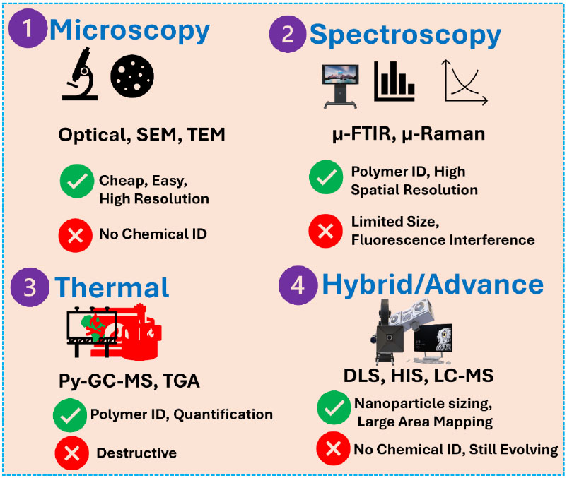
The applicability of any analytical method depends on the objective of the research. For instance, destructive mass-based methods are also suitable for quantifying only the mass of the polymer (regardless of particle number, shape, or size). For detailed information, the nondestructive particle-based methods can be applied. Thompson et al.,1966 employed a technique integrating pyrolysis and chromatography for detecting vehicle tire abrasion products in the roadway. De Leeuw et al., 1986 first reported the presence of polystyrene as an anthropogenic pollutant in sediment and soil analyzed by Py-GC/MS. Subsequently, Fabbri and colleagues employed Py-GC/MS for analysis of PS, PVC, polybutadiene, styrene-butadiene random and block, and poly(acrylonitrile-styrene-co-butadiene) in coastal sediments (Fabbri et al., 1998; Fabbri et al., 2000). Recently, Lin et al., 2020, explored the applicability of matrix-assisted laser desorption/ionization time-of-flight mass spectrometry (MALDI-ToF/MS) for the analysis of MNPs. Currently,
Fourier Transform Infrared (FTIR) spectroscopy and Raman micro-spectroscopy are widely employed for MNPs analysis because these methods enable the determination of polymer type, together with the number, size, and shape of MNPs (Koelmans et al., 2019; Schymanski et al., 2021). FTIR spectroscopy was employed to identify MPs from marine samples in the 2004 publication by Thompson et al. 2004, which also introduced the term “microplastics.”
Since then, this approach has proven to be effective for identifying, quantifying, and characterizing MP contamination in aquatic and terrestrial environments and continues to be the most relevant in MP research globally (Renner et al., 2018). IR spectroscopy is a nondestructive method based on the examination of molecular vibrations triggered by radiation absorption in the electromagnetic spectrum's mid-infrared (MIR) region (4000−400 cm−1).
Attenuated Total Reflection (ATR) can be employed for the effective study of MPs. Primpke et al., 2020, claimed that this method has been employed in 58% of IR research, particularly for larger particles due to its high cost-effectiveness. ATR-FTIR is frequently used to identify visually presorted particles (sizes larger than 200-500 μm) (Cabernard et al., 2018; Käppler et al., 2016) and to characterize weathered MP because it is easy to obtain information on changed particle surfaces caused by age (additional OH, C-O, and COOH). Additionally, ATR-FTIR can be used effectively to distinguish synthetic from natural (micro)fibers (Dris et al., 2018). Micro-FTIR spectroscopy (μ-FTIR), which combines an optical microscope with an FTIR spectrometer, is the most useful FTIR-based technique for the investigation of (MP) particles smaller than 500 μm. In addition to FPA μ-FTIR systems, an alternative method, Laser Direct Infrared (LDIR) analysis, appears to have high potential for the rapid, automated identification and quantification of MP particles. This was first reported recently by Scircle et al. 2020. LDIR has been employed for the analysis of MP particles >20 μm in soil and aquatic environments (river and marine water) (Scircle et al., 2020; Mughini-Gras et al., 2021). The light source, a patented Quantum Cascade Laser (QCL), is the main innovative feature.
Raman spectroscopy is a nondestructive analytical technique that is being employed more and more to analyze small microplastics in various environmental samples, such as fresh and marine water, sediments, biota, drinking water, beverages, and food (Missawi et al., 2020; Karami et al., 2017). Raman spectroscopy has recently been shown to be useful for analyzing nanoplastics (Sobhani et al., 2020). The interference caused by fluorescence, which can be caused by inorganic (such as dust particles and clay minerals), organic (such as humic substances), and (micro)biological impurities in the matrix as well as some additives (pigments), is a significant drawback of Raman spectroscopy, particularly when analyzing MPs in environmental samples (Araujo et al., 2018). The large number of spectra (several thousand) produced by automated Raman measurements necessitates time-efficient and reliable processing of spectra (baseline correction, smoothing, normalization) and their assignment (Cowger et al., 2020).
Despite significant advances in analytical techniques, the detection and characterization of nanoplastics remain technically challenging. Several methodological limitations hinder accurate identification, quantification, and characterization of nanoscale plastic particles in environmental and biological matrices. The major analytical challenges and potential future solutions are summarized in Table 6.
| S. No. | Analytical Challenge | Description of the Problem | Impact on Nanoplastic Research | Potential Future Solutions |
|---|---|---|---|---|
| 1. | Detection of ultrafine nanoplastics (<10 nm) | Most current spectroscopic and microscopic techniques lack the spatial resolution and sensitivity required to detect particles below 10 nm. | This leads to significant underestimation of nanoplastic abundance and environmental exposure levels. | Development of ultra-high-resolution imaging techniques and advanced spectroscopic tools capable of nanoscale polymer identification. |
| 2. | Low environmental concentrations | Nanoplastics are often present at extremely low concentrations in environmental and biological samples. | Difficulty in accurate quantification and increased risk of analytical uncertainty. | Improved sample pre-concentration methods and highly sensitive detection techniques. |
| 3. | Particle aggregation and transformation | Nanoplastics tend to aggregate or interact with natural organic matter and minerals in environmental matrices. | Alters particle size distribution and complicates accurate characterization. | Development of improved sample preparation and separation techniques to maintain particle integrity. |
| 4. | Distinguishing nanoplastics from natural nanoparticles | Environmental samples contain abundant natural colloids and organic particles with similar sizes and morphologies. | High risk of misidentification and analytical artifacts. | Integration of multi-technique analytical approaches combining spectroscopy, microscopy, and chemical analysis. |
| 5. | Lack of standardized analytical protocols | Sampling, extraction, and analytical procedures vary widely among studies. | Limited comparability of results across studies and regions. | Establishment of internationally standardized protocols and reference materials for nanoplastic analysis. |
| 6. | Sample preparation artifacts | Filtration, digestion, and extraction procedures may fragment plastics or introduce contamination. | Potential overestimation or mischaracterization of particle size distribution. | Development of gentle extraction protocols and contamination control strategies. |
| Limited polymer-specific identification at nanoscale | Many nanoparticle detection methods provide size information but lack chemical specificity. | Difficulty in confirming polymer composition of detected nanoparticles. | Coupling high-resolution imaging with advanced spectroscopic techniques such as Raman, FTIR, or mass spectrometry. |
6. CHALLENGES AND MITIGATION STRATEGIES FOR MICRO- AND NANOPLASTIC POLLUTION AND TOXICITY IN THE ENVIRONMENT AND BIOLOGICAL SYSTEMS
MNPs interact with almost all terrestrial and aquatic organisms at multiple life stages. Due to their heterogeneous nature and surface properties, they pose a growing global challenge for all organisms (Kochanek et al., 2025). The greatest challenge is the detection of nanoplastics in the complex biological and environmental matrices. Most current detection and quantification tools lack the resolution required to precisely assess nanoplastics smaller than 10 nm in these complex matrices. This limitation prevents accurate assessment of the risks posed by MNPs to exposed organisms (Wright and Kelly 2017). In addition, various physical, biological, and chemical fragmentation processes are continuously generating MNPs by weathering, photolysis, hydrolysis, wind abrasion, oxidative degradation, and biodegradation. This clearly shows that only source reduction cannot overcome the challenges posed by MNPs. The heterogeneous nature of MNPs further complicates cross-study comparisons and assessment of toxicity caused by them in diverse ecosystems. Additionally, they adsorb and transport numerous heavy metals, organic waste, microbes, and other hazardous pollutants across diverse ecosystems, thereby causing toxicity. Due to their small size (especially less than 10 nm), they cross diverse biological barriers such as intestinal epithelium, alveolar epithelium, blood-brain barrier, blood-testis barrier, and placental barrier. This leads to the accumulation of MNPs in diverse organs of the organism, such as the intestine, lung, brain, liver, testis, kidney, heart, and placenta. Their accumulation causes various inflammations and disorders like oxidative stress, disruption of mitochondrial functioning, weakening of membrane integrity, and disruption of functions of the liver, kidney, heart, spleen, pancreas, and reproductive system of the organisms (Xie et al., 2025).
Mitigation of these adverse effects of MNPs is essential and requires integrated strategies. These strategies involve source reduction through improved waste management and reduced plastic use; the adoption of advanced MNP removal technologies such as coagulation, filtration, and advanced oxidation processes; and the use of biological and biochemical solutions (Priya et al., 2022). There is an urgent need to adopt Extended Producer Responsibility (EPR) frameworks and promote numerous biodegradable alternatives. Several nature-inspired processes like nano-enabled catalytic processes and enzymatic /microbial degradation processes can overcome these challenges but require further optimization for mass applications. In addition, harmonized analytical standards, risk-based policymaking, and robust monitoring systems can aid in overcoming these challenges. Numerous regulatory policies, such as single-use plastic bans, mandatory use of filters for industrial and textile effluents, and bans on the use of microbeads, have demonstrated remarkable reductions in MNPs and their adverse effects (Malinowski et al., 2023). Increased public awareness programs, circular economy frameworks, and advanced waste management techniques can further assist in overcoming these challenges. In this section, we have presented various challenges, accounting, mitigation strategies, and representative examples of MNPs and summarized them in table format (Table 7).
| S. No. | Challenge | Account | Mitigation Strategies | Representative Examples | Ref. |
|---|---|---|---|---|---|
| 1. | Fragmentation into nanoplastics | Macroplastics progressively degrade into MNPs that are more bioavailable, mobile, and difficult to remove. | Depletion of plastic production and usage; Implementation of anti-littering policies; stabilization of polymers to prevent embrittlement. | Bans on single-use plastics; usage of stabilizers to extend material durability. | (Kochanek et al., 2025) |
| 2. | Environmental persistence | MNPs exhibit extremely slow degradation rates, leading to long-term accumulation in terrestrial and aquatic environments. | Development of biodegradable polymers; improvement of waste collection and recycling infrastructure. | Use of PLA and PHA; enhanced municipal recycling programs. | (Wright and Kelly 2017) |
| 3. | Complex environmental transport pathways | MNPs disperse via atmospheric deposition, wastewater effluents, surface runoff, and trophic transfer. | Advanced filtration systems; adoption of nature-based solutions; improved stormwater management. | Membrane filtration in WWTPs; vegetative buffer strips. | (Priya et al., 2022) |
| 4. | Adsorption of hazardous chemicals and pathogens | MNPs act as vectors for organic pollutants, heavy metals, and microbial pathogens, enhancing their environmental persistence and toxicity. | Advanced treatment processes; engineering of low-adsorption polymer materials; removal of co-contaminants. | Ozonation, AOPs, and activated carbon treatment | (Xie et al., 2025) |
| 5. | Bioaccumulation and trophic transfer | MNPs accumulate in plants, animals, and humans. | Developing dietary exposure guidelines; establishing monitoring programs; reducing agricultural plastic applications | Routine surveillance of MNPs in crops and seafood | (Malinowski et al., 2023) |
| 6. | Ecotoxicity and human health impacts | Exposure to MNPs induces oxidative stress, genotoxicity, inflammatory responses, and developmental toxicity. | Standardized toxicological testing; establishment of exposure limits, and Comprehensive risk assessment frameworks. | Formulation of reference doses and occupational safety thresholds. | (Yee et al., 2021) |
| 7. | Agricultural contamination | Plastic mulch films, contaminated fertilizers, and sewage sludge introduce MNPs into agricultural soils. | Pre-treatment of sludge before land applications, regulation of mulch films; adoption of biodegradable alternatives. | Bio-based mulch films, EU regulations on sludge application. | (Sintim and Flury) |
| 8. | Wastewater treatment inefficiencies | Conventional WWTPs have limited efficiency in removing particles <1 µm. | Adoption of rapid sand filtration, membrane bioreactors, and tertiary treatments such as AOPs. | Reverse Osmosis (RO) systems and Ultrafiltration (UF). | Monira et al., 2023 |
| 9. | Limitations in detection and quantification | Detection of microplastics and especially nanoplastics is challenging due to small size and complex sample matrices. | Harmonization of global monitoring protocols; establishment of high-resolution analytical techniques. | Nanoparticle tracking. Raman spectroscopy, µ-FTIR, Py-GC-MS. | (Ośko et al., 2025) |
| 10. | Limited public awareness | High dependency on single-use plastics and inadequate waste segregation drives environmental release. | Incentives for recycling; public education campaigns; implementation of Extended Producer Responsibility (EPR) policies. | Community recycling initiatives; plastic buy-back programs. | (Heidbreder et al., 2023) |
| 11. | Regulatory gaps | Lack of harmonized regulations and exposure thresholds hampers risk management. | Standardized risk assessment methodologies; Development of international regulatory frameworks. | EU and UNEP initiatives on exposure standards. | (Bearth et al., 2025) |
7. CONCLUSION AND FUTURE PERSPECTIVES
This review article amalgamates current knowledge on the sources, physicochemical properties, environmental transport, bioaccumulation, and biological impacts of MNPs. The article highlights the environmental fate and toxicity of MNPs, which are strongly governed by their physicochemical characteristics, including shape, size, surface charge, morphology, and polymer composition. These characteristics regulate environmental mobility, contaminant adsorption capacity, biological uptake efficacy, and various toxicity pathways.
Owing to their extremely small size and high surface reactivity, nanoplastics exhibit greater environmental mobility and bioavailability than microplastics. They can easily penetrate many biological barriers such as pulmonary, placental, intestinal, and blood-brain barriers. This leads to systemic distribution and accumulation of MNPs in organs such as the liver, kidney, lungs, and brain. Experimental evidence shows that MNP exposure can trigger oxidative stress, inflammation, mitochondrial dysfunction, genotoxicity, and programmed cell death in both animal and plant systems. In plants, nanoplastics interfere with nutrient transport, stomatal function, metabolic activity, and gene expression, ultimately affecting plant growth and productivity.
Human exposure to microplastics has been estimated at 103-105 particles per day via ingestion and 102-104 particles per day via inhalation. However, these values are highly uncertain due to methodological variability and a lack of measurements of nanoplastics. Translating these exposure estimates into risk-relevant measures remains difficult since there is no agreement on the appropriate dose descriptor (particle mass, number, and surface area), and internal dose data is extremely restricted. A screening-level quantitative risk assessment shows that microplastic concentrations in sediment and freshwater systems approach/exceed Predicted No-Effect Concentration (PNEC) limits under high-exposure scenarios. Freshwater PNEC estimates using species sensitivity distributions (2.61 particles L−1; 0.003-0.011 mg L−1) (Morales-Espinoza et al., 2025) are consistent with upper-bound environmental concentrations reported in damaged systems (Morales-Espinoza et al., 2025; Pandey et al., 2023). Sediment concentrations of 105 particles kg−1 (Patra et al., 2024) are comparable to projected effect thresholds, indicating a localized ecological danger. However, the significant diversity in Risk Quotients (RQ) among locations (RQ = 0.000008-22.06) demonstrates the importance of spatial heterogeneity and methodological uncertainty (Morales-Espinoza et al., 2025). Furthermore, the lack of rigorous nanoplastic quantification results in a systematic underestimate of overall exposure and risk (Bae et al., 2025).
Nevertheless, the current understanding of MNP behavior and toxicity remains fragmented due to limitations in analytical techniques, inconsistent experimental conditions, and insufficient long-term exposure studies. Therefore, the development of a comprehensive understanding of the structure-activity relationships controlling the environmental fate and biological impacts of MNPs remains a crucial challenge for environmental science and toxicology.
To address these limitations, the following research priorities should be addressed:
(1) Development of standardized analytical methods for NPs (nanoplastics) detection. Sensitive and validated techniques are required to accurately detect, quantify, and characterize NPs nanoplastics in complex environmental and biological matrices.
(2) Understanding structure-activity relationships of MNPs. Future studies should systematically investigate how physicochemical parameters such as size, surface charge, morphology, polymer type, and aging influence environmental transport, bioavailability, and toxicity.
(3) Assessment of long-term and low-dose exposure effects. Most existing studies focus on short-term laboratory exposure. Long-term and environmentally relevant exposure studies are necessary to evaluate chronic ecological and human health risks.
(4) Mechanistic understanding of MNP interactions with biological systems. Integrated omics approaches, including transcriptomics, proteomics, and metabolomics, should be employed to clarify molecular mechanisms underlying MNP-induced toxicity in animals, plants, and microorganisms.
(5) Integration of environmental transport models with ecological risk assessment. Future research should link environmental distribution, trophic transfer, and bioaccumulation processes with toxicological outcomes to develop more accurate risk assessment frameworks and inform regulatory policies.
To improve the global understanding of MNPs pollution and develop effective strategies to mitigate environmental and health impacts, the aforementioned scientific challenges should be addressed.
AUTHORS’ CONTRIBUTIONS
The authors confirm their contribution to the paper as follows: M.K.G. and A.G.: Data analysis, interpretation, writing the manuscript, and supervision; M.K., C.A., M.P., D.D., and I.M.: Methodology, supervision, and manuscript drafting. All authors reviewed the results and approved the final version of the manuscript.
ACKNOWLEDGEMENTS
Declared none.

